Methods and Devices of early diagnosis of corneal diseases
The cornea is the outmost tissue of the human eye and represents its main objective lens. Any disease of the tissue may strongly compromises human vision. The corneal tissue consists of five layers which, from the outside to the inside, are represented by the epithelium, the Bowman’s membrane, the stroma, the Descemet membrane and the endothelium. The epithelium consists of several cell layers; it is able to regenerate thanks to the activity of the limbal stem cells. The stroma represent the core of the tissue and is mainly composed of collagen.
The pathologies of the corneal tissue represent the third global cause of serious vision loss worldwide. Trauma, infections or genetic diseases can compromise tissue integrity and may cause loss of corneal transparency. Despite continuous advances in ophthalmic technologies, there is not currently an ophthalmic instrument that may resolve the main component of the cornea, the collagen!
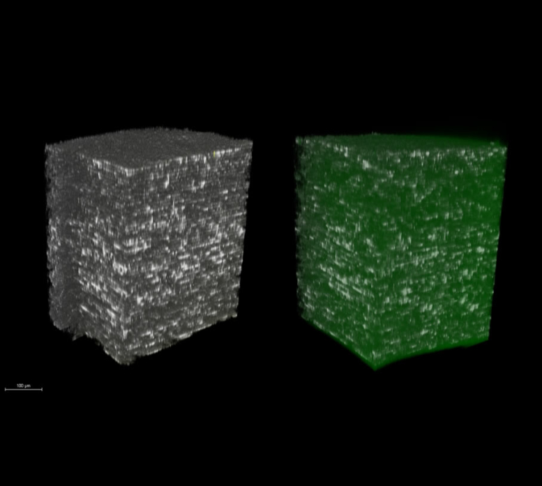
Vision Engineering Italy is working on a novel highly innovative solution for the early diagnosis of corneal diseases. The study is conducted in collaboration with the most important research centers in biophotonics and high resolution microscopy accross Europe. The technology is based on two-photon non-linear microscopy.
Publications
- Lombardo G, Micali NL, Villari V, Leone N, Rusciano D, Serrao S, Lombardo M. Assessment of stromal riboflavin concentration-depth profile in nanotechnology-based transepithelial corneal cross-linking. J Cataract Refract Surg 2017;43(5):680-686.
- Lombardo M, Parekh M, Serrao S, Ruzza A, Ferrari S, Lombardo G. Two-photon optical microscopy imaging of endothelial keratoplasty grafts. Graefe’s Arch Clin Exp Ophthalmol 2017; 255(3):575-582
- Lombardo M, Micali N, Villari V, Serrao S, Lombardo G All-optical method to assess stromal concentration of riboflavin in conventional and accelerated UV-A irradiation of the human cornea. Invest Opthalmol Vis Sci 2016; 57(2): 476-483.
- Lombardo M, Merino D, Loza-Alvarez P, Lombardo G. Translational label-free nonlinear imaging biomarkers to classify the human corneal microstructure. Biomed Opt Express 2015; 8;6(8):2803-18.
- Labate C, Lombardo M, De Santo MP, Dias J, Ziebarth N, Lombardo G. Multiscale investigation of the depth-dependent mechanical anisotropy of the human corneal stroma. Invest Ophthalmol Vis Sci 2015; 56: 4053-4060.
- Alizadeh M, Merino D, Lombardo G, Lombardo M, Mencucci R, Ghotbi M, Alvarez P. Identifying crossing collagen fibers in human corneal tissues using pSHG images. Biomed Opt Expr 2019 Jul 10;10(8):3875-3888.

